
Calcinosis in Systemic Sclerosis
Systemic sclerosis (or scleroderma) is characterized by many skin changes, including calcinosis. In this article, we will discuss what is calcinosis, how it is diagnosed and available treatments.
WHAT IS CALCINOSIS?
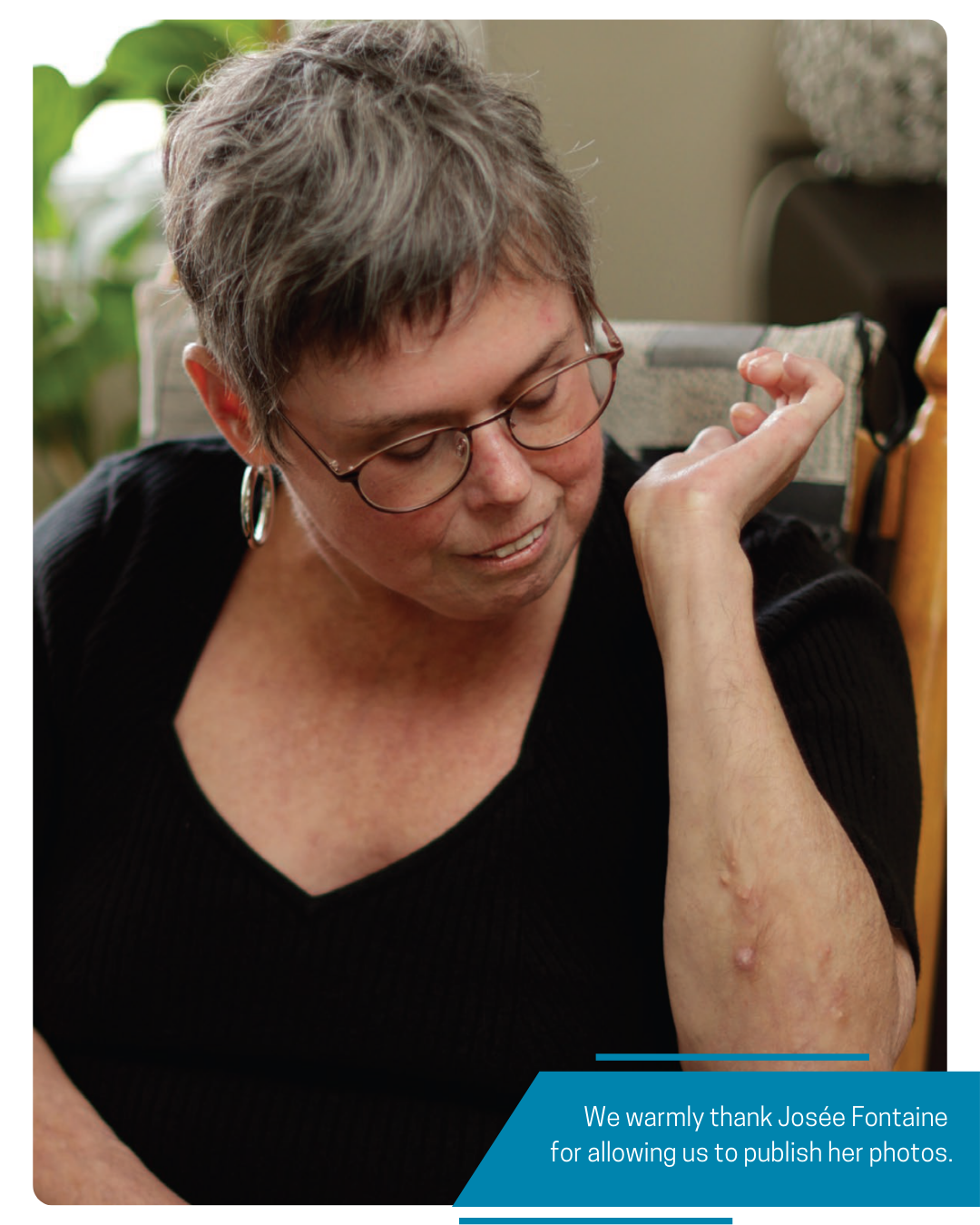
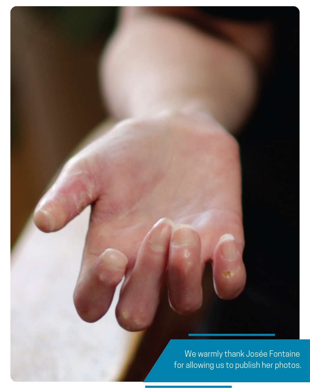
Calcinosis is an accumulation of calcium in the skin and surrounding tissues. It can affect up to 20-40% of people with scleroderma, with a similar rate between limited and diffuse scleroderma. It often occurs on the hands, forearms, elbows, and knees, despite normal calcium levels in the blood. Small lesions may go unnoticed, but may also cause pain, interfere with joint function, or be complicated by ulcers, infections, or nerve compression. Unfortunately, few cases of calcinosis improve spontaneously with time, with most lesions remaining stable or worsening after 1 year.
 |
WHY DOES CALCINOSIS DEVELOP, AND WHICH SCLERODERMA PATIENTS ARE MOST AT RISK? We still do not completely understand why calcinosis develops. Studies have found that people with scleroderma who are men, who have ulcers on their fingers, who have osteoporosis, or internal organ involvement of scleroderma (especially lung fibrosis) are at higher risk for calcinosis. Some autoantibodies have also been associated with calcinosis, including anti-centromere, anti-PM-Scl and anti-RNA-polymerase-3 autoantibodies. Other potential factors include longer disease duration, poor blood circulation, and trauma.
HOW IS CALCINOSIS DIAGNOSED? It is often felt by the physician on physical exam and can be confirmed by plain radiographs. Other ways to identify calcinosis include ultrasound, computed tomography and magnetic resonance imaging.
|
|
HOW DO WE TREAT CALCINOSIS?
Calcinosis remains a challenging aspect of scleroderma to manage, as there is no cure or highly effective treatment. We generally do not treat calcinosis that does not bother a patient. At present, no medical therapies have proven to be efficacious in large randomized controlled trials.
General Measures
Generally, it is recommended to avoid injuries and ensure good blood flow to prevent new calcinosis, as these are thought to play a role in calcinosis development. This includes smoking cessation, avoiding exposure to cold and stress, and managing Raynaud and ulcers on the fingers. If a wound develops, proper wound care is important, and antibiotics should be given for nonhealing and infected ulcers. If there is an open wound, warm soaks can help squeeze out calcinosis and may help prevent infections.
Medical Therapies
Sodium thiosulfate is thought to improve calcinosis because it binds to calcium. A systematic review that looked at 40 studies (including 1 small randomized controlled trial) found that the topical form of this medication improved calcinosis in over 80% of patients over an average of 5 months. Topical sodium thiosulfate needs to be prepared by a pharmacist in a mixture with either Vaseline or zinc oxide.
Possible side effects include skin irritation, allergy to zinc and pain with application. Sodium thiosulfate can also be injected directly into calcinosis lesions, usually by a dermatologist. A case series of 5 scleroderma patients showed improvement or complete resolution of calcinosis with weekly injections for 4 weeks. A common side effect is a burning sensation in the area of the injection site.
Several medications have also been shown to improve calcinosis in small studies. Two pill form medications, colchicine and minocycline, may decrease the inflammatory aspect of calcinosis, when there is associated pain, redness, warmth and swelling, as shown in small studies involving less than 10 patients each. One of the side effects of colchicine is diarrhea, while the side effects of minocycline include nausea, dizziness, and a blueblack discoloration of calcinotic lesions. Other medications, such as diltiazem, bisphosphonates, rituximab, and intravenous immunoglobulins, have been reported to help calcinosis in small studies, but with no larger or higher quality studies to support these observations.
Finally, neem oil with Hypericum plant extract is an herbal therapy that is often used in wound care. A pilot study of 21 scleroderma patients with open calcinotic wounds followed over 40 days found that neem oil helped complete healing in 45% of patients, perhaps by softening and facilitating excision of calcium deposits. This would need to be further supported in larger studies.
 |
Interventions Surgery to remove calcinosis can be an effective treatment and is considered for lesions that are affecting hand or joint function, compressing a nerve, or causing severe discomfort. Complications include recurrence in 15%, delayed wound healing in 13%, and wound infection in 10% of cases. Extracorporeal shockwave therapy uses acoustic shock waves to break apart and destroy calcinosis. It is performed by physiotherapists who have received the appropriate training. It can help relieve pain associated with calcinotic lesions that are not amenable to surgery. Several prospective studies have found improvements in pain and size of calcinotic lesions. It often involves 3 to 5 sessions done every 7 to 10 days.
In Summary Calcinosis is a feature of scleroderma that is common but unfortunately difficult to treat. Important preventative measures include avoiding trauma and ensuring good blood circulation such as through smoking cessation, cold avoidance, proper management of Raynaud, and treating ulcers. For calcinotic lesions that are symptomatic, topical or injected sodium thiosulfate may be helpful. Surgery may be considered for lesions that significantly affect function and quality of life. More research is needed to understand the cause and optimal treatment of calcinosis.
|
|

REFERENCES:
1. Nowaczyk J, Zawistowski M, Fiedor P. Local, non-systemic, and minimally invasive therapies for calcinosis cutis: a systematic review. Arch Dermatol Res 2022;314(6):515-25. doi: 10.1007/s00403-021-02264-5 [published Online First: 20210624]
2. Elahmar H, Feldman BM, Johnson SR. Management of Calcinosis Cutis in Rheumatic Diseases. J Rheumatol 2022;49(9):980-89. doi: 10.3899/jrheum.211393 [published Online First: 20220515]
3. Davuluri S, Lood C, Chung L. Calcinosis in systemic sclerosis. Curr Opin Rheumatol 2022;34(6):319-27. doi: 10.1097/ bor.0000000000000896 [published Online First: 20220819]
April 2024